The Future in Mind

The Future in Mind

Anthony W.P. Fitzpatrick, PhD
Assistant Professor of Biochemistry and Molecular Biophysics; Principal Investigator at Columbia’s Zuckerman Institute
Proteins: New Details of Neurodegeneration Emerge
Parkinson’s. Huntington’s. ALS. These devastating neurodegenerative diseases and many more have something in common. Each has been linked to a protein in the brain that malfunctions and forms clumps, turning them from helpful to harmful.
Neuroscientist Anthony Fitzpatrick has been scrutinizing these proteins in unprecedented detail. He maps their structures in the brain, atom by atom, using a microscope developed by a Columbia Nobel laureate.
“Our technology here at the Zuckerman Institute, cryogenic electron microscopy, is the most powerful tool science has to look at protein complexes,” said Dr. Fitzpatrick.
Consider tau proteins. They stick together and form tangles that are a hallmark of Alzheimer’s and other brain diseases called tauopathies. In 2017, Dr. Fitzpatrick gave the world its first three-dimensional look at tau protein misfolded in the brain (inside brain tissue donated by a deceased patient). In the following years his team took a closer look at tau proteins and discovered a specific structural modification that causes them to stick together. More recently, the team found that tauopathies and other seemingly unrelated diseases might share a surprising underlying cause: not tau, but a different misfolded protein that had escaped notice—until Dr. Fitzpatrick’s team put it under their microscope.
With this structural information in hand, scientists can think about designing new treatments that target these malformed proteins.
“When you know the structure of a protein, you can ask better questions about what happens to the protein in a cell and how to control it,” said Dr. Fitzpatrick. “Our research offers a way to target the root causes of a disease rather than its symptoms.”
An analysis of cryo-electron microscope images revealed how the enormous protein megalin (LRP2) changes shape (A. Fitzpatrick, A Beenken, L. Shapiro/Columbia’s Zuckerman Institute).
Photo credit: A. Fitzpatrick, A Beenken, L. Shapiro/Columbia’s Zuckerman Institute
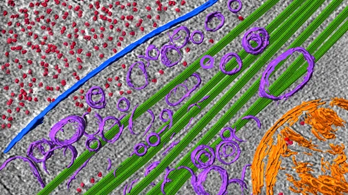
Cryo-Electron tomography reveals the molecular architecture of the interior of a neurite. Ribosomes (red), microtubules (green), vesicles (purple), single membrane (blue), double membrane (orange).
Photo credit: Fitzpatrick lab/Columbia’s Zuckerman Institute

Proteins:
New Details of Neuro-degeneration Emerge
Parkinson’s. Huntington’s. ALS. These devastating neurodegenerative diseases and many more have something in common. Each has been linked to a protein in the brain that malfunctions and forms clumps, turning them from helpful to harmful.
Neuroscientist Anthony Fitzpatrick has been scrutinizing these proteins in unprecedented detail. He maps their structures in the brain, atom by atom, using a microscope developed by a Columbia Nobel laureate.
“Our technology here at the Zuckerman Institute, cryogenic electron microscopy, is the most powerful tool science has to look at protein complexes,” said Dr. Fitzpatrick.
Consider tau proteins. They stick together and form tangles that are a hallmark of Alzheimer’s and other brain diseases called tauopathies. In 2017, Dr. Fitzpatrick gave the world its first three-dimensional look at tau protein misfolded in the brain (inside brain tissue donated by a deceased patient). In the following years his team took a closer look at tau proteins and discovered a specific structural modification that causes them to stick together. More recently, the team found that tauopathies and other seemingly unrelated diseases might share a surprising underlying cause: not tau, but a different misfolded protein that had escaped notice—until Dr. Fitzpatrick’s team put it under their microscope.
With this structural information in hand, scientists can think about designing new treatments that target these malformed proteins.
“When you know the structure of a protein, you can ask better questions about what happens to the protein in a cell and how to control it,” said Dr. Fitzpatrick. “Our research offers a way to target the root causes of a disease rather than its symptoms.”
An analysis of cryo-electron microscope images revealed how the enormous protein megalin (LRP2) changes shape.
Photo credit: A. Fitzpatrick, A Beenken, L. Shapiro/Columbia’s Zuckerman Institute
Cryo-Electron tomography reveals the molecular architecture of the interior of a neurite. Ribosomes (red), microtubules (green), vesicles (purple), single membrane (blue), double membrane (orange).
Photo credit: Fitzpatrick lab/Columbia’s Zuckerman Institute
DOWNLOAD Anthony’S PROFILE